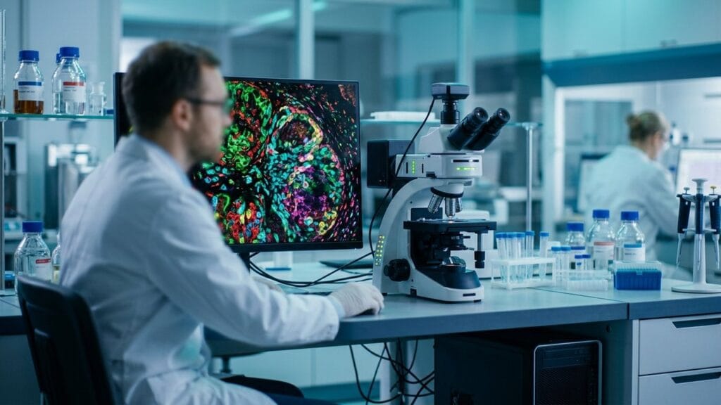
Traditional tissue analysis reads a lone marker per slide, which leaves most of the story untold. Multiplex imaging changes that by reading a whole panel in a shared pass, all within the same sample, so researchers can see how cells sit and signal together. The result is a far fuller view of a tissue than older methods ever delivered. A researcher sees the whole neighborhood instead of a narrow doorway. The tissue stays in place throughout, which keeps every measurement honest. A careful lab always keeps the staining steps identical from sample to sample. True reproducibility comes from steady habits rather than clever shortcuts.
The technique has moved from a specialty trick to a core tool in oncology, immunology, and drug discovery. Laboratories now treat it as a standard part of the toolkit. The panel grows richer as more targets join the study. Each new marker adds a layer of understanding to the same tissue. The method always rewards patience, since the setup work benefits countless samples later. This guide explains how multiplex imaging works, why it matters, and the advantages that make it indispensable in modern laboratories.
What Multiplex Imaging Does
Multiplex imaging detects a group of molecular markers in the same tissue section at the same time. Each marker carries its own label, and the microscope reads every label in the same run. The labels stay distinct because each glows in a unique color. Samples prepared well give answers that rushed samples never will. A steady hand at the bench always beats a clever plan at the desk. The images improve as the team gains practice with the platform. The output is a layered map of a sample rather than a narrow snapshot.
From a Marker to a Full Panel
Older approaches fit a basic stain per slide, which forced teams to cut serial sections and guess how they lined up. Multiplex imaging removes that guesswork, since the whole panel lives together in the tissue. The sample stays intact, and the spatial story stays truthful. What the eye finally sees is the tissue as it truly is arranged.
The Role of Spatial Context
Biology happens in place. A cell’s behavior depends on its neighbors, and multiplex imaging preserves that neighborhood. The technique shows not just what is present, but where it sits and what it touches. Position is the missing dimension that older methods quietly discarded. Training on test sections shortens the learning curve. Documenting the steps makes the work repeatable for the next study. Clear records help a colleague follow the same path.
Why Multiplex Imaging Matters
Diseases are rarely about an isolated protein. They are ecosystems of cell types that cooperate, compete, and signal. A method that sees only a hidden target misses the ecosystem entirely. The tissue is a community, and communities must be studied whole.
A Fuller View of Disease
Multiplex imaging captures the immune cells, the tumor cells, and the supporting tissue in the same frame. That combined view reveals how a tumor escapes attack or how a therapy reshapes the surrounding landscape. No older staining method can match that depth of context. The field keeps advancing as new dyes and platforms appear. Researchers share panels and protocols so the whole field moves faster. Open methods spread the benefit beyond a lab’s own walls. No older staining method can match that depth.
Speed and Sample Use
Tissue samples are small and hard to replace. Reading a full panel from a shared section conserves material that serial sections would exhaust. The approach also compresses a long workflow into a shorter task, since a compact run replaces a stack of separate stains. Fewer steps mean fewer places for error to hide. A well-run study stands up to review and repetition. The technique grows more useful as the tools grow more capable. Teams that plan ahead get the most from every tissue block.
How Multiplex Imaging Works
The workflow divides into a few clean stages, and each stage builds on the last.
Labeling the Targets
Antibodies tagged with distinct dyes find and bind their targets in the tissue. The dyes are chosen so that no two colors bleed into each other. The binding can happen together or in rounds, depending on the platform. Each dye glows in its own color, which keeps the markers separate.
Reading the Signal
A specialized microscope illuminates the sample and records every color channel. The instrument resolves the channels so that neighboring dyes do not blur into each other, which preserves accuracy as the panel grows. A steady instrument keeps the readout dependable. Careful storage keeps samples ready for a future panel. The method turns archived tissue into a lasting resource. Every laboratory adapts the workflow to its own questions.
The Analysis Step
Software then turns the images into numbers. It counts cells, measures their spacing, and maps which types sit beside which. The result is a table that a statistician can trust. That analysis step is where the raw picture becomes a biological finding.
Groundbreaking Advantages
Eight advantages explain why multiplex imaging keeps spreading across laboratories, and each advantage addresses a real limit of the older methods. Together they add up to a better way of seeing. The same platform serves studies across different disease areas. Each lab writes its own protocol notes and shares them freely. Shared notes spare a newcomer months of trial and error.
One Sample, A Full Panel
A shared tissue section now yields a full panel of markers. That conservation preserves samples and gives every experiment a broader result.
True Spatial Relationships
Because the markers share a section, the positions are real rather than reconstructed. What sits beside what is a fact of the tissue, not a guess. The community improves the method through open exchange. A thoughtful panel question beats a broad shotgun approach. Specific questions lead to specific, useful answers. Start with the question, then choose the markers that answer it. Neighboring cells are truly neighbors, which makes interaction studies reliable.
Deeper Cell Identification
Combined markers define cell types that a basic stain cannot. A cell that looks alike by a narrow marker can be sorted correctly by its full profile.
Fewer Experimental Variables
A compact run with a steady instrument and a steady sample removes the drift that creeps in across serial sections. The comparison between markers becomes cleaner and more honest. Honest data is the foundation of every serious study.
Richer Data Per Sample
Each sample now contributes a set of measurements instead of just a couple. The same tissue that earlier supported a narrow conclusion now supports a layered result.
Faster Answers
A full panel in a compact pass shortens the time from question to result. Teams reach insight sooner without cutting corners on depth. Speed and depth rarely travel together, yet here they do.
Better Use of Limited Material
Small biopsies and archived blocks go further when a shared section carries the whole analysis. The method protects the samples that cannot be replaced.
A Bridge to AI Analysis
The layered, quantitative output suits modern analysis tools well. Machines thrive on the rich maps the method delivers. Machines learn patterns from the rich maps that multiplex imaging produces, which opens the door to predictive insights.
Where Multiplex Imaging Is Used
The technique has found a home in varied fields, and each field leans on a different strength of the method.
Oncology
Cancer research uses multiplex imaging to map tumors and their immune surroundings in a combined view. The result guides treatment choices and reveals why some patients respond while others do not. That kind of clarity was impossible with older tools.
Immunology
Immune studies need to see numerous cell types together. Multiplex imaging shows the full cast of an immune response and how the players arrange themselves in tissue.
Neuroscience
Brain tissue holds complex circuits that span a range of cell types. The method maps those types side by side, which helps untangle how healthy and diseased brains differ. The wiring of the brain slowly comes into view.
Drug Discovery
Early testing benefits from the depth multiplex imaging provides. A candidate compound can be judged by how it reshapes the tissue ecosystem, not just by a narrow readout. The tissue itself becomes the scoreboard.
Practical Considerations
Adopting multiplex imaging involves a modest set of decisions, and thinking them through keeps a project on track.
Panel Design
Pick markers that answer the question rather than every marker available. A focused panel reads cleanly, while an overstuffed panel blurs the signal and the story. Less often really is more in panel design.
Platform Choice
Platforms differ in how large a panel they resolve and how they bind the targets. Match the platform to the tissue type and the question, and test with a small run beforehand.
Analysis Discipline
The analysis is where results live or fall. Keep the scoring consistent, document every step, and let a colleague review the pipeline before trusting the numbers. A fresh pair of eyes catches what the busy maker misses. The tissue guides the panel, not the other way around. A good pilot run reveals problems while they are still easy to fix. Fixing a panel early beats redoing a full study later.
How the Results Are Read
The method produces a stack of images, and reading them well is a skill of its own. The raw colors become insight only after careful interpretation.
Compare the Channels
Each channel marks a different target, and comparing them side by side reveals where the markers overlap. That overlap often points to the most important biology in the sample.
Count and Measure
Software counts the cells and measures the gaps between them. These numbers turn a colorful picture into a testable result that can be shared and repeated.
Look for Patterns
The final step is pattern finding. A cluster here, a gradient there, a border of immune cells around a tumor; these patterns are the goal of the whole workflow.
Choosing the Right Platform
Not every platform fits every study, and the choice shapes what the data can later say.
The Main Detection Styles
Some platforms bind all targets in a whole round, while others cycle the labels in steps. The whole-round approach is faster, and the stepped approach reaches wider panels.
Match to the Tissue
Dense tissues and delicate samples each favor a different setup. A trial run on a test section settles most questions before the real experiment begins.
Think About the Analysis Early
The best platform choice is the approach that pairs cleanly with the analysis tools the team already trusts. Planning both together avoids a painful mismatch later.
Common Mistakes
A frequent error is building a panel too broad, which muddies the readout and wastes sample. Another slip is skipping a control run on a new platform, which lets an instrument quirk hide inside real data. A further error is treating the map as the finish line rather than the start, since the biological question still needs answering after the image is made. The image is the beginning, never the end.
Conclusion
Multiplex imaging has redefined what a shared tissue sample can say. By reading a full panel in an intact section, it reveals the spatial and cellular detail that older methods flattened away. The advantages, from sample conservation to richer data, explain its rapid rise across research and drug discovery. For any team that needs to see the whole story in a clean view, multiplex imaging is the tool that finally makes it possible.
Frequently Asked Questions
What is multiplex imaging?
It is a technique that detects a group of molecular markers in a tissue sample at the same time, preserving their spatial layout.
How does multiplex imaging differ from standard staining?
Standard staining reads a basic marker per slide, while multiplex imaging reads a full panel in a section, with positions intact.
Which fields use multiplex imaging most?
Oncology, immunology, neuroscience, and drug discovery rely on it to map cell types and their surroundings.
How many markers can multiplex imaging detect?
The count depends on the platform and chemistry, ranging from a handful to over a hundred markers in a section.
Does multiplex imaging work with archived samples?
Yes, it often works on stored tissue blocks, which extends its reach to valuable historical collections.
Is special software needed for multiplex imaging?
Yes, analysis software quantifies the channels and maps cell relationships, turning the images into usable data.